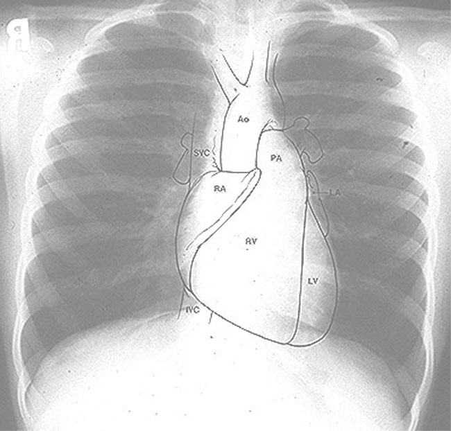
Normal Chest X-ray
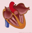
Left Atrial Dilatation
 |
| Prominence of left atrial appendage (straightening or convex) with double atrial shadow |
Right Atrial Dilation
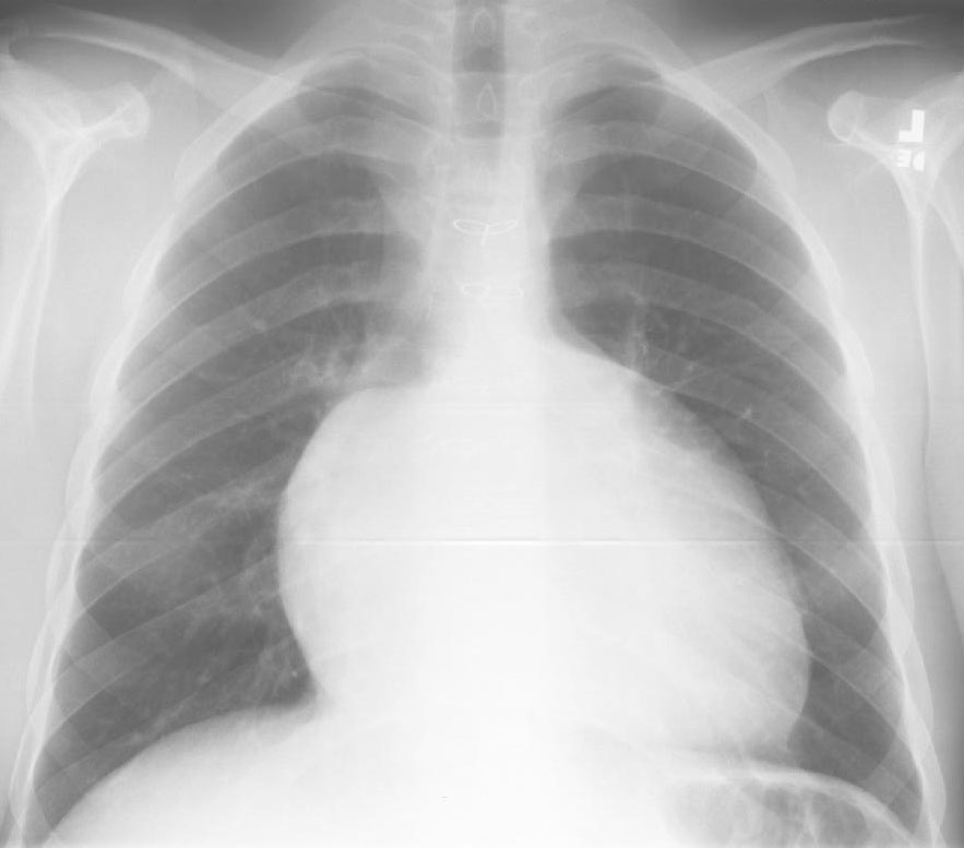 |
| Right boarder of the heart projecting into the right lower lung field. |
Electrocardiogram Results :~
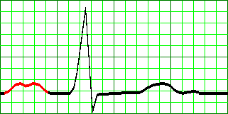
a. An electrocardiographic criterion of left atrial enlargement is a
P wave (>0,12sec) that is too wide or inverted in lead II and V1 :
P mitrale
*normal PR interval 0,12-0,20 sec
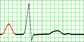
b. An electrocardiographic criterion of right atrial enlargement is a
P wave (>0,28sec) that is high amplitude in lead II and V1 : P pulmonal
c. An electrocardiographic criterion of biatrial enlargement is a
P wave that is too tall and too wide in lead II.
The conclusion for atrial investigations :)~
 |
| Left Atrial Enlargement (LAE) |
 |
| Right Atrial Enlargement (RAE) |